
Ear
Bone-anchored hearing implants and Contact Forte: how to choose
A bone-anchored hearing implant carries sound to the inner ear through vibration of the skull bone. It is indicated for malformations of the ear, conductive or mixed hearing loss that does not suit a conventional hearing aid, and deafness in one ear. The three implantable models available differ in size and in the degree of loss they cover. Contact Forte is the non-surgical option, with less powerful sound.
Written by Profa. Dra. Aline Bittencourt, CRM-MA 7562 Medically reviewed by Profa. Dra. Aline Bittencourt, CRM-MA 7562 Published on
For some people, a conventional hearing aid is not a good option: the ear canal never formed, chronic otitis keeps the ear from staying dry, or the deafness is in one ear only. In these cases there is another route for sound to reach the inner ear: the bone of the skull. This is how bone-anchored hearing implants work.
How does sound reach the inner ear?
Sound has to reach the cochlea, the organ of hearing, in the inner ear. It can get there by two routes:
- Through the air: sound enters the ear canal and makes the eardrum vibrate. The vibration passes through the ossicles, the three tiny bones of the middle ear (malleus, incus and stapes), and reaches the cochlea.
- Through the skull bones: the vibration crosses the bone of the head and goes straight to the cochlea, bypassing the canal, the eardrum and the ossicles.
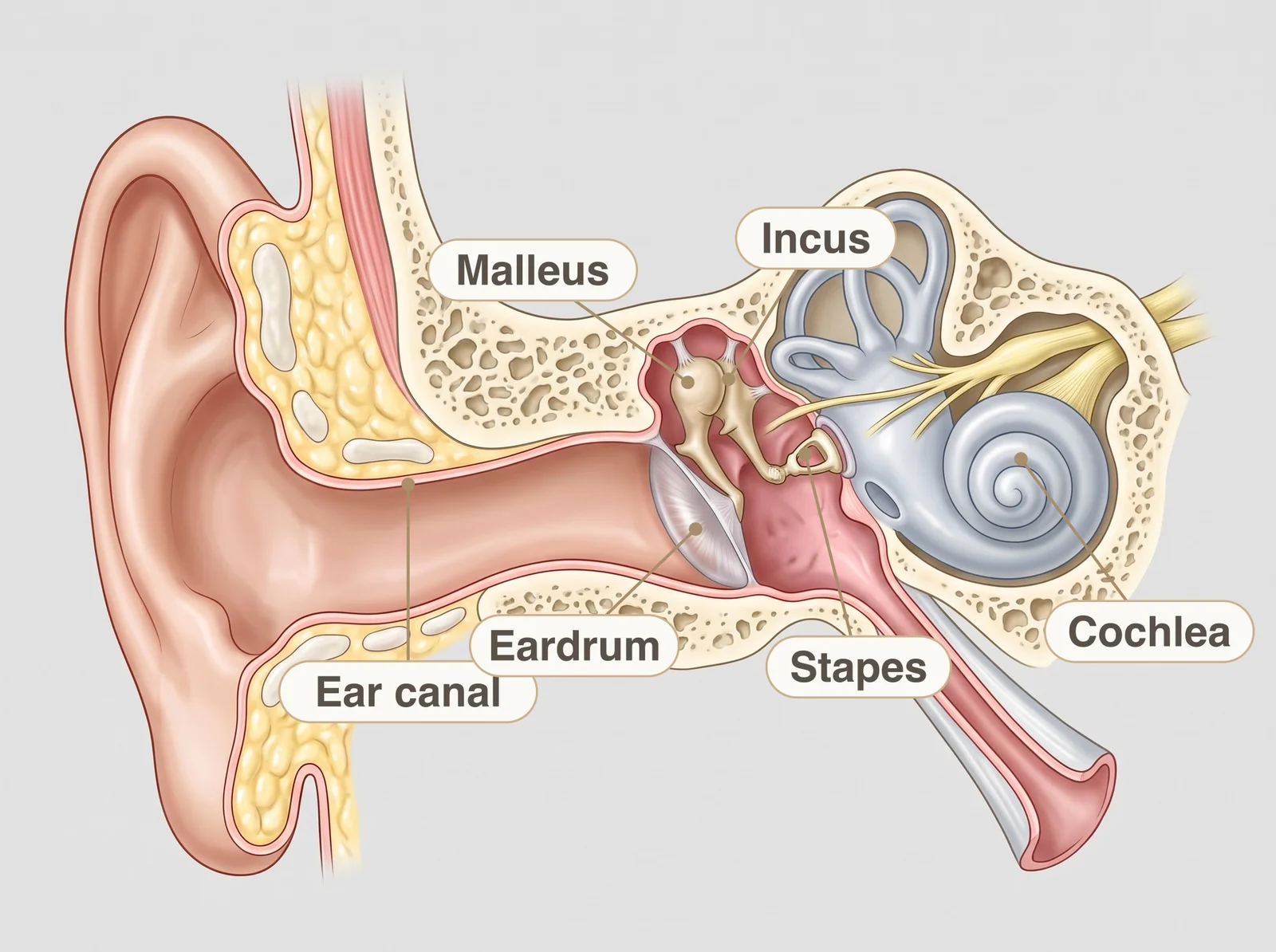
A bone-anchored implant uses the second route. That is why it can help when the problem lies in the canal, the eardrum or the ossicles, and also in deafness in one ear.
What are the parts of the implant?
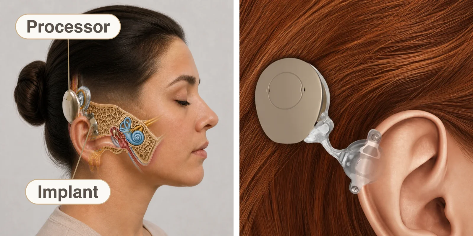
- Internal part: the implant, placed in the bone behind the ear during surgery. It is completely covered by skin, with nothing passing through it, and does not need replacing unless it fails.
- External part: the sound processor, held on the skin by a magnet. It picks up sounds and turns them into vibration, which the implant carries through the bone to the cochlea.
Who can use one?
A bone-anchored implant may be indicated for people who:
- Were born with a malformation of the outer ear or ear canal that prevents the use of a conventional hearing aid.
- Have conductive or mixed hearing loss and do not get on with a conventional aid, as in chronic otitis media or after mastoid surgery.
- Have single-sided deafness: the implant is placed on the deaf side and carries sound, through the bone, to the good ear on the other side.
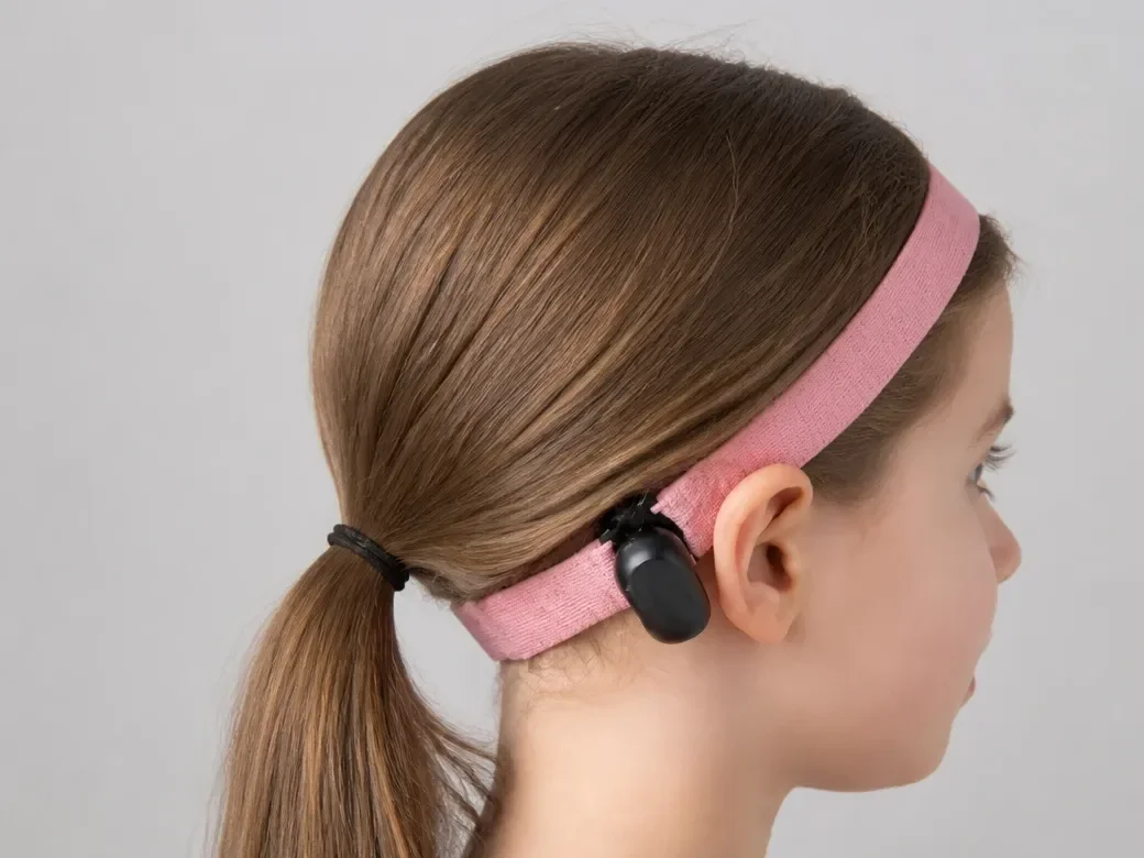
Surgery is indicated from 5 years of age. Before that, the skull bone is still thin and growing, which does not allow the implant to be fixed safely. Until then, a child can wear a processor on a soft headband, without surgery, so as not to go without hearing stimulation.
Which models are there?
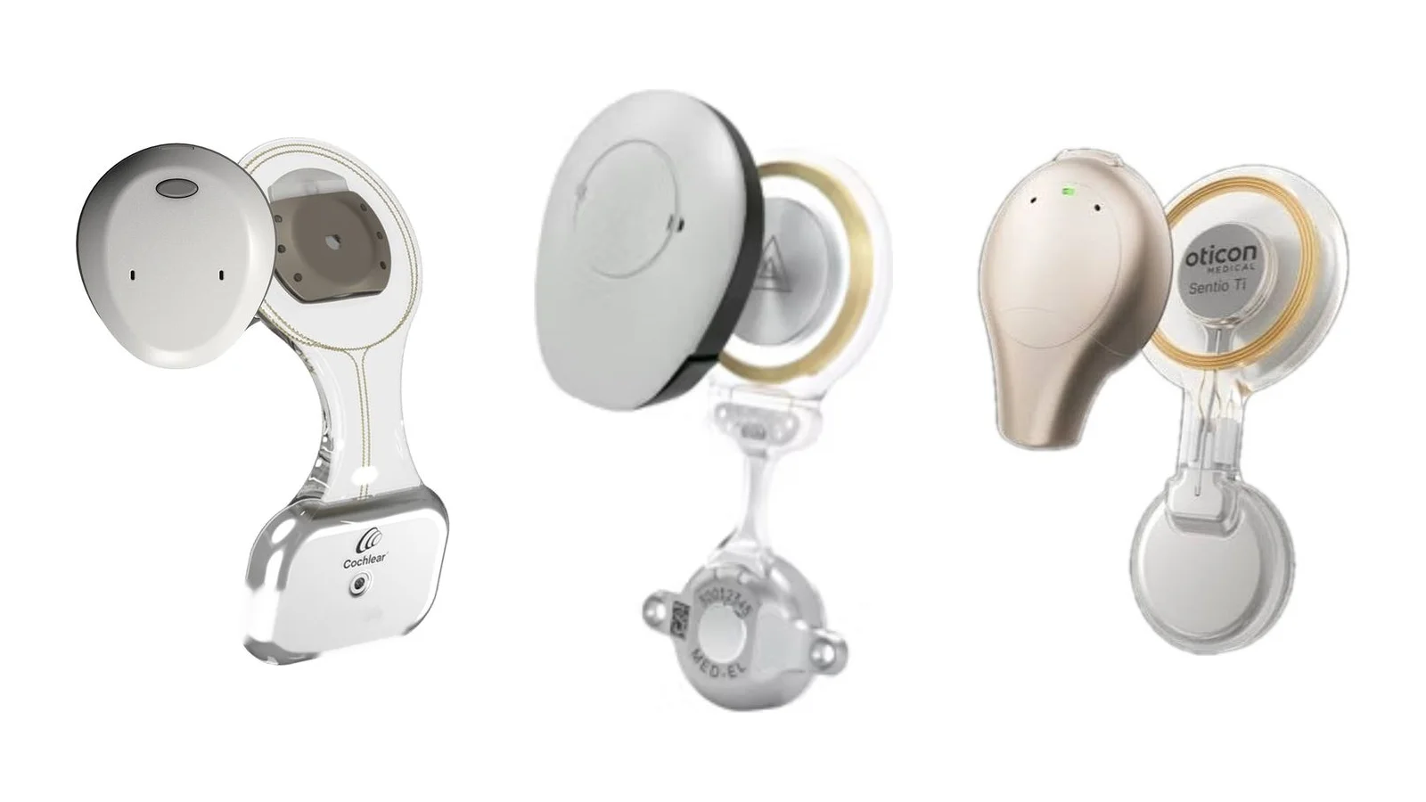
Three models are available in Brazil, all imported: Osia, Bonebridge and Sentio. In all three, the implant sits under the skin and the processor attaches to it by a magnet, which is why they are called transcutaneous implants. They differ in size and in the degree of loss they cover.
The implant of each model
| Osia | Bonebridge | Sentio | |
|---|---|---|---|
| Manufacturer | Cochlear | MED-EL | Oticon Medical |
| Implant size | 7.2 × 3.1 cm | 6.4 × 2.8 cm | 6.0 × 3.0 cm |
| Bone conduction loss* | Up to 55 dB | Up to 45 dB | Up to 45 dB |
| MRI** | Up to 1.5 tesla; 3 tesla with the magnet removed | Up to 1.5 tesla | Up to 1.5 tesla |
* Measured on audiometry: the higher the number, the greater the inner-ear loss the model can cover. ** Always following the manufacturer’s safety conditions.
The sound processor of each model
| Osia | Bonebridge | Sentio | |
|---|---|---|---|
| Processor | Osia 2 | SAMBA 2 | Sentio 1 Mini |
| Size | 3.6 × 3.2 cm | 3.6 × 3.0 cm | 3.9 × 3.0 cm |
| Colours | 5: black, sand beige, slate grey, silver grey and brown | 7 in hair tones, plus 10 decorated covers | 6: beige, terracotta, brown, steel grey, black and silver |
| Power | Disposable 675 battery | Disposable 675 battery | Disposable 675 battery |
Measurements are approximate, according to the manufacturers. No model is better than another in the abstract: the right one depends on the tests, the anatomy of the bone, the likely need for MRI and each person’s daily life.
The non-surgical option: Contact Forte
Contact Forte is a bone conduction device that does not need surgery. Resting behind the ear, it transmits the vibration of sound through the skin to the inner ear.
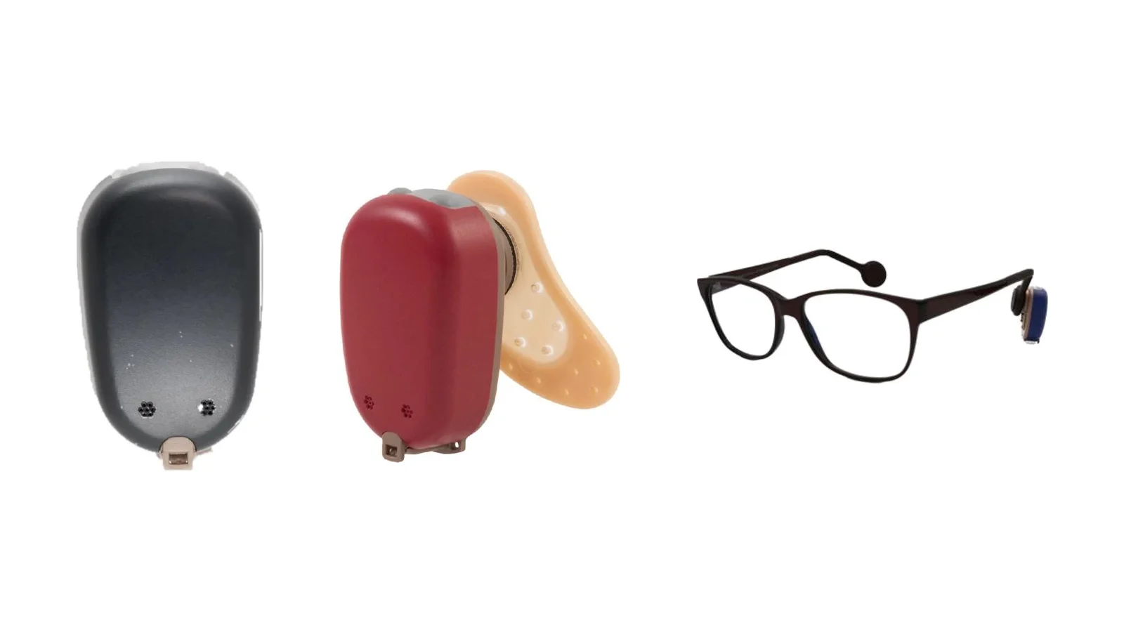
Ways to wear it:
- Soft headband, in several sizes, including for babies.
- Adhesive on the skin behind the ear, in front of the hairline.
- Glasses, with an adapter on the arm.
- Cap, which holds the device behind the ear.
It may be indicated:
- For children under 5.
- For people who cannot have, or do not want, surgery.
- To try the sound before deciding on surgery.
- For a period, while waiting for surgery.
Implant or Contact Forte?
| Bone-anchored implant | Contact Forte | |
|---|---|---|
| Surgery | Yes, under general anaesthesia | No |
| Age | From 5 years | Any age, including babies |
| How it is held | Processor held by a magnet over the implant | Headband, adhesive, glasses or cap |
| Path of the vibration | Straight through the bone, from the implant | Crosses the skin before reaching the bone |
| Strength of sound | Greater | Lower, because the skin dampens the vibration |
| Most common use | Long-term use | Young children, a trial before surgery, or people who will not have surgery |
Which tests are done before surgery?
- Audiometry: measures hearing in each ear and shows whether the implant can help.
- Trial with the processor: done by the audiologist, with the device on a headband, to assess the benefit before surgery.
- CT scan of the temporal bones: shows the thickness and quality of the bone behind the ear and helps plan the best site for the implant.
- Blood count, clotting tests and assessment by a cardiologist and anaesthetist, to confirm that general health allows surgery.
How is the operation done?
The operation is performed in hospital under general anaesthesia and takes about an hour and a half on average. The patient is asleep throughout and feels no pain.
- An incision behind the ear, a small cut in the skin.
- Preparing the bone, where a small space is made to receive the implant.
- Fixing the implant to the bone. Over time it bonds with the bone and becomes firm, which is called osseointegration.
- Closure and dressing: the skin is stitched over the implant, and the head is wrapped in a bandage that stays on for 3 days.
Patients usually go home the same day or the next.
What happens afterwards?
- Recovery: mild pain, swelling and numbness around the scar are expected. The bandage is removed 3 days after surgery and the stitches at 14 days, at the clinic.
- Activation: done by the audiologist from 20 days after surgery, once the implant is firm in the bone. Until then, hearing does not change. At first, sounds may seem different; adaptation is gradual and individual.
- Implant card: a manufacturer’s document that identifies the model and serial number and states the safe conditions for MRI. It should be kept for life and shown before imaging tests and at metal detectors in banks and airports.
What are the risks?
Like any operation, implant surgery has risks, which are discussed before the decision:
- Failure of the implant to integrate, so that it does not bond with the bone and another operation is needed.
- Infection, which may require removal of the implant.
- Haematoma, a collection of blood under the skin that may need draining.
- Skin reaction under the magnet, with pain, redness or, more rarely, a sore at the site of the processor. It may require adjusting the magnet strength, local treatment or further surgery.
Lifelong care
The implant has a magnet and metal parts, and some situations need attention:
- MRI: all three models allow scans up to 1.5 tesla, following the conditions on the implant card. The magnet can leave a dark area on images near the implant, so head scans should be discussed beforehand. CT and ultrasound have no restriction.
- Metal detectors: they may set off the alarm. Simply show the implant card.
- Other operations and treatments: always tell the surgeon, dentist and anaesthetist. Monopolar electrosurgery should not be used on the head and neck, and treatments with electric or magnetic fields near the implant need prior assessment.
- Water and sport: remove the processor for showering, swimming and the sea, unless the model is waterproof, and for contact sports.
What is important to know before deciding?
The implant does not restore normal hearing, but it can improve the ability to hear and understand sound, and results vary from person to person. The processor needs daily care, and regular follow-up with the ENT and the audiologist helps look after the skin, the implant and the processor settings.
Indications, assessment and the choice of model are covered on the page on bone-anchored hearing implants.